Introduction:
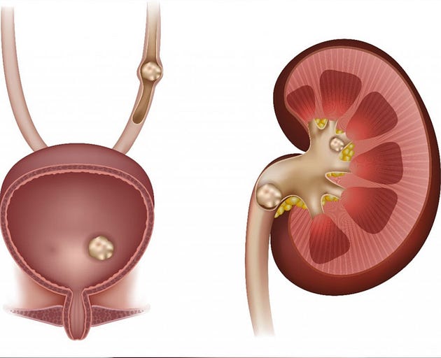
When it comes to kidney stones, prompt diagnosis is crucial for effective treatment and management. However, identifying these small, hard mineral deposits within the kidneys or urinary tract can be challenging without the right tools and techniques. In this blog post, we’ll explore the various methods healthcare providers use to diagnose kidney stones, shedding light on the diagnostic process and the technologies involved.

Clinical Evaluation:
Diagnosing kidney stones typically begins with a thorough clinical evaluation, where the healthcare provider collects information about the patient’s medical history, symptoms, and risk factors. Understanding the patient’s symptoms, such as sudden and severe pain in the back, abdomen, or groin, along with associated signs like nausea, vomiting, and blood in the urine, can provide valuable clues suggestive of kidney stones.
Diagnostic Imaging:
Once a suspicion of kidney stones is raised based on clinical evaluation, diagnostic imaging tests are often employed to confirm the diagnosis and assess the size, location, and number of stones present. Several imaging modalities may be utilized for this purpose:
1. X-ray:
X-ray imaging is commonly used to detect the presence of kidney stones, particularly those composed of calcium. However, not all types of kidney stones are visible on standard X-rays, limiting its sensitivity for certain stone types like uric acid or cystine stones.
2. Computed Tomography (CT) Scan:
CT scans are highly effective in diagnosing kidney stones, offering detailed cross-sectional images of the urinary tract. CT scans can accurately visualize even small stones and provide information about their location, size, and potential complications such as obstruction or infection. While CT scans are sensitive and reliable, they do expose patients to ionizing radiation, which may be a consideration, especially for pregnant women and children.
3. Ultrasound:
Ultrasound imaging uses sound waves to create images of the kidneys and urinary tract. While not as sensitive as CT scans for detecting small stones, ultrasound can still be valuable, particularly in situations where minimizing radiation exposure is a priority, such as during pregnancy or in pediatric patients. Additionally, ultrasound is useful for evaluating complications such as hydronephrosis (fluid buildup in the kidneys) associated with kidney stones.
Urine Tests:
In addition to imaging studies, urine tests may be conducted to analyze the composition of the urine and identify factors that contribute to stone formation. These tests can help determine the presence of substances like calcium, oxalate, uric acid, and cystine, which are common components of kidney stones. Urine tests may also reveal signs of urinary tract infections or metabolic abnormalities that increase the risk of stone formation.
Conclusion:
Diagnosing kidney stones requires a comprehensive approach that integrates clinical evaluation, imaging studies, and urine tests. By combining these diagnostic modalities, healthcare providers can accurately identify the presence of kidney stones, assess their characteristics, and formulate appropriate treatment plans tailored to the individual patient’s needs. Whether it’s through advanced imaging technologies like CT scans or non-invasive methods like ultrasound, early and accurate diagnosis is essential for guiding timely intervention and preventing complications associated with kidney stones.
Comments
Post a Comment