
What are the common diagnostic methods for detecting kidney stones?

 Detecting kidney stones typically involves a combination of clinical evaluation, imaging techniques, and sometimes laboratory tests. 
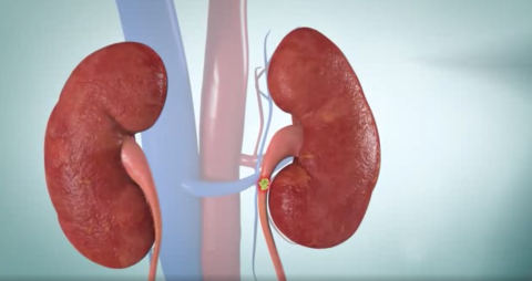
Here are the common diagnostic methods used:

  1. Symptom Assessment and Medical History: The first step is often a detailed discussion about the patient’s symptoms. Symptoms such as severe pain in the side and back, blood in urine, nausea, and vomiting are indicative and help narrow down the diagnosis.
  2. Physical Examination: A physical examination can provide additional clues, such as tenderness in the abdomen or lower back.
  3. Imaging Tests:
  • CT Scan (Computed Tomography): This is the preferred imaging method for detecting kidney stones due to its high sensitivity and specificity. It can accurately locate the stones, determine their size, and assess complications like obstruction.
  • Ultrasound: Particularly useful in pregnant patients or when avoiding radiation exposure is a concern. Ultrasound can detect larger stones but may miss smaller ones or those located in certain parts of the urinary tract.
  • X-ray: Used less frequently now due to its limitations in detecting all types of stones, but can still be used in certain cases, especially if a CT scan is not available.

4. Laboratory Tests:

  • Urinalysis: Detects blood in the urine (hematuria) and can also identify signs of infection or abnormal pH levels that may contribute to stone formation.
  • Blood Tests: These may be done to assess kidney function and evaluate for conditions that predispose to stone formation, such as hyperparathyroidism.

5. 24-Hour Urine Collection: In some cases, a 24-hour urine collection may be requested to analyze the levels of stone-forming substances like calcium, oxalate, uric acid, and citrate. This helps in planning preventive strategies to reduce the risk of future stone formation.

6. Intravenous Pyelogram (IVP): While less commonly used now due to the availability of CT scans, an IVP involves injecting a contrast dye into a vein, which is filtered by the kidneys and excreted into the urinary tract. X-rays are then taken to visualize the flow of urine and any obstructions caused by stones.

Each diagnostic method has its advantages and limitations, and the choice of which method to use depends on factors such as the patient’s symptoms, medical history, availability of imaging equipment, and the clinical judgment of the healthcare provider. Early and accurate diagnosis is crucial for timely management and prevention of complications associated with kidney stones.

Comments

Popular posts from this blog

Can you explain the different types of minimally invasive surgeries available in urology?

 Minimally invasive surgeries in urology are designed to reduce the physical trauma of traditional surgical methods while still effectively treating urological conditions. These techniques generally involve smaller incisions, less pain, quicker recovery times, and minimal scarring.  Here’s a look at the various types of minimally invasive surgeries commonly used in urology: 1. Laparoscopic Surgery Laparoscopic surgery , also known as keyhole surgery, involves making small incisions (typically 0.5–1 cm) through which a camera (laparoscope) and specialized surgical instruments are inserted. The camera provides real-time images of the internal organs, allowing the surgeon to perform the procedure with precision. Applications: Kidney Surgery : Removal of kidney tumors or partial nephrectomy (removal of part of the kidney). Adrenal Surgery : Removal of adrenal gland tumors. Bladder Surgery : Treatment of bladder tumors or diverticula. Advantages: Less postoperative pain. Shorter ho...

Risks and Complications Associated with Kidney Transplant Surgery

  Kidney transplant surgery is a life-changing procedure that offers many patients with end-stage renal disease (ESRD) a renewed chance for a healthier and more active life. However, like any major surgery, it carries certain risks and potential complications. Understanding these risks can help patients and their families make informed decisions and better prepare for the post-transplant period. 1. Surgical Risks a. Infection: As with any major surgery, kidney transplant recipients are at risk for infections. The surgery itself can introduce bacteria, and the use of immunosuppressive medications to prevent organ rejection weakens the body’s natural defense mechanisms, making infections more likely. b. Bleeding: Significant bleeding during or after the surgery is a potential risk. Although surgeons take extensive measures to minimize this risk, some patients may require blood transfusions or additional procedures to manage bleeding complications. c. Anesthesia Complications:...

What is a kidney transplant, and who is a candidate for this procedure?

 A kidney transplant is a surgical procedure where a healthy kidney from a living or deceased donor is implanted into a recipient whose kidneys treatment have failed or are functioning at a critically low level. This procedure provides a long-term solution to kidney failure, offering recipients a chance to regain near-normal kidney function and significantly improve their quality of life. Candidates for Kidney Transplant: End-Stage Kidney Disease (ESKD) : Individuals whose kidneys have lost nearly all their ability to function, leading to dangerous levels of waste products and fluids in the body, are candidates for a kidney transplant. Chronic Kidney Disease (CKD) : In some cases, individuals with advanced CKD that is progressing rapidly may be considered for a transplant before reaching ESKD to improve long-term outcomes. Health Status : Candidates must be in relatively good health apart from their kidney condition. They should be free from active infections, cancers that could af...